Best Oral Cancer Surgeon in Varanasi | Dr. Atul Kumar Rai

Discover why Dr. Atul Kumar Rai is recognized as the best oral cancer surgeon in Varanasi. Read this comprehensive guide on identifying early mouth cancer signs, diagnostic biopsies, advanced surgical resections, microvascular reconstruction, and recovery protocols at Star Hospital.

Understanding Oral Cavity Malignancies: A Complete Guide by the Best Oral Cancer Surgeon in Varanasi

Oral cavity and head and neck cancers represent a significant healthcare challenge across Eastern Uttar Pradesh, Bihar, and Jharkhand. Driven predominantly by chronic exposure to chewable tobacco, gutkha, paan masala, betel nut, and smoking, thousands of individuals suffer from non-healing mouth ulcers, restricted mouth opening (Oral Submucous Fibrosis or OSMF), and malignant tissue growths every year.

When facing an oral cancer diagnosis, securing early intervention from an expert surgical oncologist is the single most critical factor determining long-term survival, complete tumor clearance, and functional rehabilitation.

If you or a loved one are searching for the best oral cancer surgeon in Varanasi, this comprehensive guide by Dr. Atul Kumar Rai outlines everything you need to know—from recognizing early warning signs to navigating advanced surgical resections, microvascular reconstruction, and post-operative recovery.
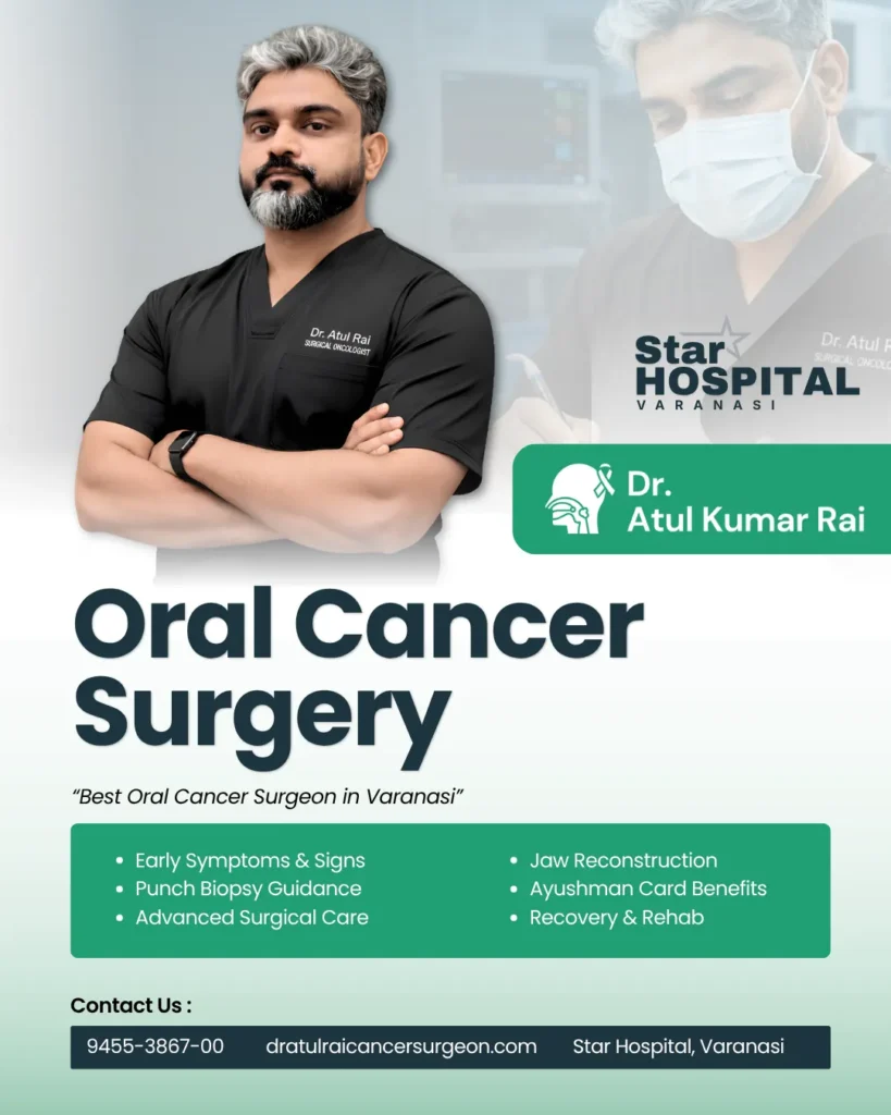
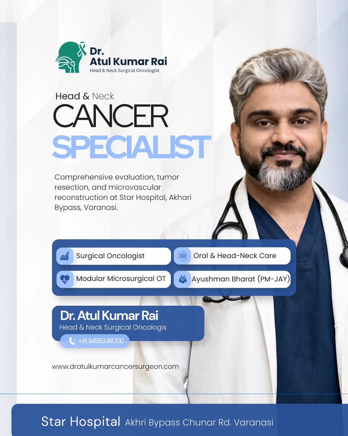
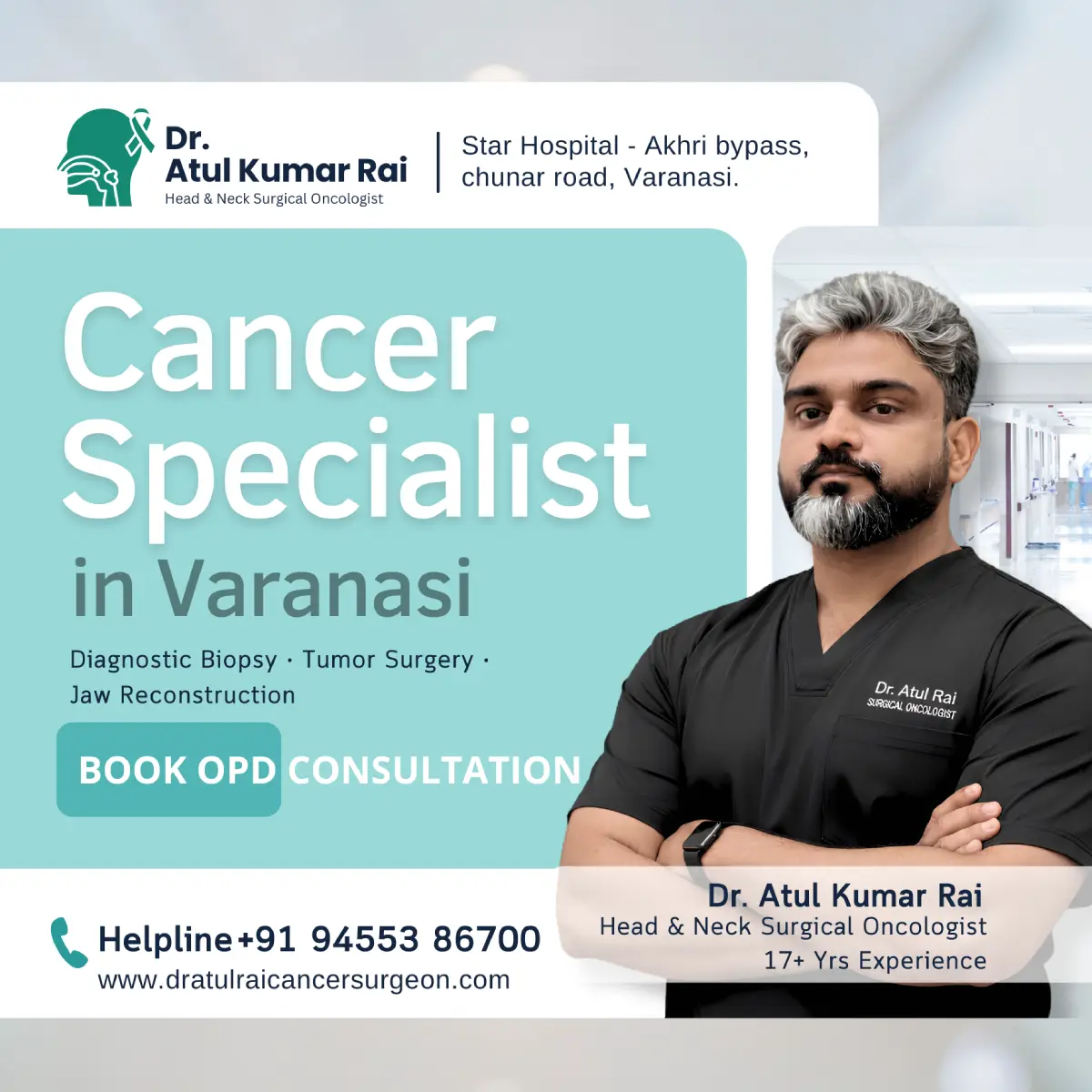
Dr. Atul Kumar Rai Best Oral Cancer Surgeon in Varanasi performing consultation

The Rising Burden of Oral Cancer in Eastern Uttar Pradesh

The Purvanchal region, with Varanasi serving as its central medical hub, sees a high incidence of oral cavity malignancies. The lining of the mouth—known clinically as the oral mucosa—is highly sensitive to regular chemical irritation. Continuous friction from sharp teeth, tobacco consumption, and alcohol usage triggers chronic inflammation, which over time alters cellular DNA, leading to precancerous lesions and overt invasive carcinomas.

The primary sub-sites within the oral cavity susceptible to cancer include:

  • Buccal Mucosa: The inner lining of the cheek (the most frequent site of tobacco-related cancer in India).

  • Anterior Two-Thirds of the Tongue: Malignancies on the sides or underside of the tongue.

  • Floor of the Mouth: The tissue beneath the tongue.

  • Gums & Alveolar Ridge: The mucosal tissue covering the upper and lower jawbones.

  • Hard Palate: The bony roof of the front of the mouth.

  • Retromolar Trigone: The small area located behind the wisdom teeth.

  • Lips: Particularly lower lip carcinomas linked to pipe smoking or direct sun exposure.

Early Warning Signs: When Should You Consult an Oral Cancer Specialist?

One of the greatest challenges in treating mouth cancer is delayed presentation. Many patients mistake early malignant lesions for routine aphthous ulcers, dental trauma, or vitamin deficiencies, allowing the disease to advance from stage I to stage III or IV before seeking specialist care.

+———————————————————————–+
| EARLY VS. ADVANCED SYMPTOMS |
+———————————————————————–+
| EARLY SIGNS (Act Immediately) | ADVANCED SIGNS (Urgent Onco-Care) |
+———————————–+———————————–+
| * Non-healing ulcer (>14 days) | * Severe ear pain (referred pain) |
| * Red or white patches | * Difficulty swallowing or chewing|
| * Unexplained bleeding in mouth | * Unexplained loosening of teeth |
| * Painless lump in cheek or neck | * Restricted tongue movement |
+———————————————————————–+

1. The 14-Day Rule for Mouth Ulcers

A routine mouth ulcer caused by stress or accidental biting typically heals within 7 to 10 days. Any sore, erosion, or ulcer in the oral cavity that persists for more than 14 days despite routine anti-inflammatory or antiseptic treatment must be evaluated by a head and neck surgical oncologist for a diagnostic biopsy.

2. Precancerous Conditions: Leukoplakia, Erythroplakia & OSMF

Before an invasive cancer forms, the oral lining often displays precancerous changes:

  • Leukoplakia: A prominent white patch on the cheek, tongue, or gums that cannot be scraped off.

  • Erythroplakia: A smooth, velvety red patch that carries a significantly higher risk of malignant transformation than leukoplakia.

  • Oral Submucous Fibrosis (OSMF): Characterized by progressive stiffness of the cheek mucosa, burning sensation while eating spicy food, and severe restriction in mouth opening (inability to insert 3 or 4 fingers vertically).

3. Painless Neck Lumps

Oral cancer frequently spreads to the regional cervical lymph nodes in the neck before metastasizing to distant organs. A painless, firm, or non-tender lump in the upper neck is often an early indicator that an oral cavity lesion has metastasized regionally.

Diagnostic Evaluation: How Dr. Atul Kumar Rai Diagnoses Oral Cancer

An accurate, swift diagnostic protocol forms the bedrock of successful cancer treatment. When you visit Dr. Atul Kumar Rai at Star Hospital, Varanasi, your diagnostic evaluation follows a structured, multi-step process:

[Clinical Examination] ──> [Diagnostic Biopsy / FNAC] ──> [Cross-Sectional Imaging (CT/MRI)] ──> [Staging & Surgical Plan]

1. Detailed Clinical & Endoscopic Assessment

The oncologist conducts a systematic palpation of the oral cavity, assessing the depth, mobility, and margins of the lesion, alongside a comprehensive clinical palpation of all neck node levels (Levels I through V).

2. Diagnostic Punch Biopsy

A biopsy is the ultimate definitive gold standard for confirming cancer. Under local anesthesia, a tiny tissue sample (2 to 3 mm) is taken from the edge of the ulcer or mass and sent for histopathological examination (HPE). Pathologists analyze the tissue under a microscope to identify the exact tumor type—most commonly Squamous Cell Carcinoma (SCC)—and its differentiation grade.

3. Cross-Sectional Staging Imaging

To determine the depth of invasion (DOI) and check if the tumor has eroded into adjacent structures like the mandible (lower jawbone) or maxilla (upper jawbone), advanced imaging is performed:

  • Contrast-Enhanced CT (CECT) Scan: Excellent for assessing bone erosion and cervical lymph node involvement.

  • Magnetic Resonance Imaging (MRI): Provides superior soft-tissue resolution, vital for evaluating tongue cancers and soft tissue depth.

  • PET-CT Scan: Indicated in advanced stages to rule out distant organ spread (lungs, liver, bones).

Why Surgery is the Primary Curative Treatment for Oral Cancer

While treatment modalities for cancer include chemotherapy, radiation therapy, and immunotherapy, surgical resection remains the primary, definitive curative treatment for cancers of the oral cavity.

+———————————————————————–+
| THE TRIAD OF ONCOLOGICAL SURGERY |
+———————————————————————–+
| 1. COMPLETE RESECTION | 2. NECK NODE DISSECTION | 3. RECONSTRUCTION |
| Achieving clear (R0) | Clearing regional lymph | Restoring form, |
| margins around tumor | nodes to prevent return | speech & swallowing|
+———————————————————————–+

As the best oral cancer surgeon in Varanasi, Dr. Atul Kumar Rai follows a strict three-phase surgical doctrine:

Phase 1: Wide Local Excision with Clear Margins (R0 Resection)

The primary objective during surgery is complete microscopic removal of all malignant tissue. Dr. Rai performs a wide local excision, removing the tumor along with a healthy safety margin of surrounding normal tissue (typically 1 to 1.5 cm around the palpable edge). Achieving “R0 or Clear Margins” is the single most important surgical factor in preventing local cancer recurrence.

Phase 2: Cervical Neck Lymph Node Dissection

Because oral cancer cells readily migrate through lymphatic channels to the neck, a neck dissection is performed simultaneously with the main tumor resection.

  • Selective Neck Dissection (SND): Performed in early-stage disease to remove lymph nodes at high risk (Levels I, II, and III) while preserving healthy muscles, nerves, and veins.

  • Modified Radical Neck Dissection (MRND): Required in node-positive cases to clear a wider array of neck tissue while retaining vital anatomical structures whenever possible.

Phase 3: Immediate Microvascular Reconstruction

Removing a sizable tumor from the cheek, tongue, or jaw creates a surgical defect that must be repaired immediately. Modern surgical oncology goes beyond tumor removal—it focuses on functional restoration.

Dr. Rai collaborates with specialized microvascular plastic surgeons to perform immediate reconstructive flap surgeries:

  • Pectoralis Major Myocutaneous (PMMC) Flap: A reliable pedicled tissue flap used to close soft tissue defects.

  • Anterolateral Thigh (ALT) Flap: A versatile soft-tissue microvascular flap ideal for reconstructing large cheek or tongue defects.

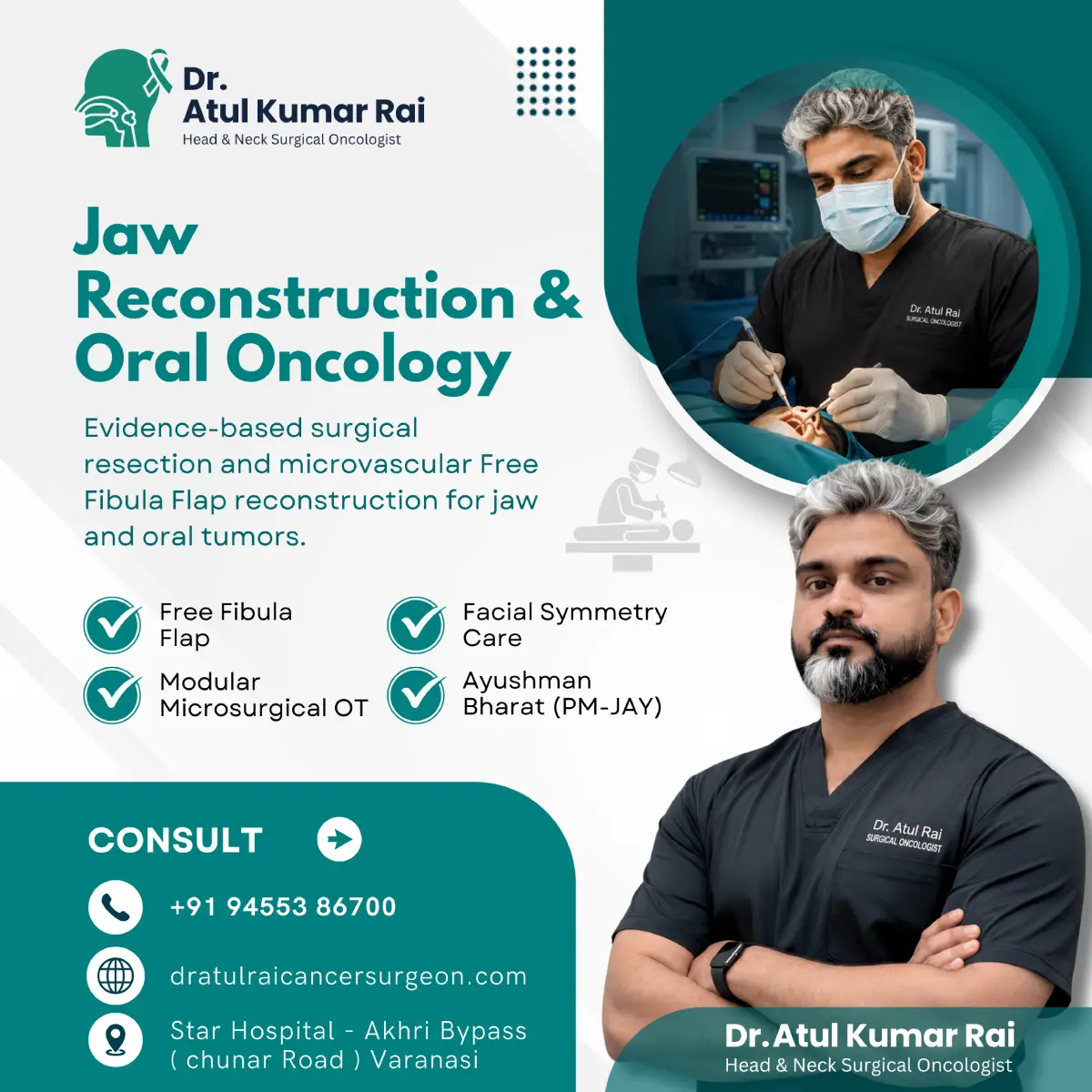
  • Free Fibula Flap: Harvesting a piece of the lower leg bone along with its blood vessels to rebuild a segment of the lower jawbone (mandible), allowing for future dental implants.

Surgical Expertise Spotlight: Dr. Atul Kumar Rai

Choosing the right surgeon for head and neck oncology directly impacts surgical safety, clearance rates, and quality of life. Dr. Atul Kumar Rai is widely regarded as a premier surgical oncologist in Eastern India due to his patient-centric approach and clinical precision.

+———————————————————————–+
| CLINICAL STANDOUTS & ADVANTAGES |
+———————————————————————–+
| * Specialized Training | High-volume expertise in head & neck surgery|
| * Function-Preserving | Nerve-sparing techniques for speech/chewing |
| * State-of-the-Art OT | Modern ICU & monitoring at Star Hospital |
| * Transparent Guidance | Clear cost structures & scheme facilitation |
+———————————————————————–+

Advanced Surgical Modalities Offered:

  • Composite Resection (Commando Operation): Complex surgery for advanced buccal mucosa lesions involving cheek excision, jawbone resection (segmental mandibulectomy), neck dissection, and flap reconstruction.

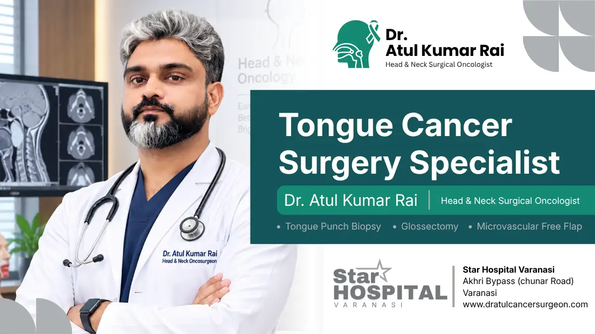
  • Partial & Total Glossectomy: Precise tongue cancer excision with submental or free flap reconstruction to retain speech clarity and swallowing function.

  • Mandibular Reconstruction: Rebuilding jaw bone integrity to maintain natural facial symmetry.

  • OSMF Surgical Release: Release of dense fibrous tissue bands in the cheek followed by skin/buccal fat pad grafting to restore normal mouth opening.

  • Thyroid & Parotid Surgery: Delicate nerve-monitoring procedures for benign and malignant head and neck glandular tumors.

What to Expect During Surgical Recovery and Rehabilitation

Recovery following oral cavity cancer surgery is a structured journey requiring skilled nursing, clinical monitoring, and multidisciplinary support.

[Days 1-3: ICU Monitoring & Airway Care]


[Days 4-7: Flap Viability Checks & Swallowing Rehab]


[Days 8-14: Wound Healing & Suture/Staple Removal]


[Weeks 3-4: Histopathology Review & Adjuvant Planning]

Oral Cancer Surgery Cost in Varanasi and Financial Access

Medical care should be accessible, transparent, and manageable for every family. The overall cost of oral cavity cancer surgery varies depending on several clinical factors:

  • Stage and Complexity: Early-stage wide excision vs. advanced composite resection.

  • Type of Reconstruction: Local pedicled flap (e.g., PMMC) vs. microvascular free tissue transfer.

  • Hospital Stay & ICU Requirements: Length of stay and specialized nursing needed.

Ayushman Bharat (PM-JAY) & Scheme Support

Dr. Atul Kumar Rai provides clinical care at Star Hospital, Varanasi, an empaneled center facilitating cashless benefits under government healthcare schemes including Ayushman Bharat Pradhan Mantri Jan Arogya Yojana (PM-JAY). Eligible patients can access life-saving surgical oncology care and hospitalization without catastrophic out-of-pocket expenses.

Preventative Advice: Protecting Yourself Against Oral Cancer

While advanced surgical oncology offers high cure rates, prevention and early detection remain the ultimate defense.

+-----------------------------------------------------------------------+
|                    PREVENTATIVE CHECKLIST FOR ORAL HEALTH             |
+-----------------------------------------------------------------------+
| [✓] Quit all forms of tobacco (Gutkha, Khaini, Cigarettes, Bidi)      |
| [✓] Limit alcohol consumption                                         |
| [✓] Schedule annual dental checks to address sharp or broken teeth     |
| [✓] Eat a fresh, antioxidant-rich diet with green vegetables & fruits |
| [✓] Perform monthly self-examinations of inner cheeks, tongue & gums  |
+-----------------------------------------------------------------------+

Frequently Asked Questions (FAQs)

Dr. Atul Kumar Rai is widely recognized as a leading surgical oncologist and oral cancer surgeon in Varanasi. He specializes in precision tumor resections (wide local excision), neck node dissections, jaw reconstruction, and microvascular free flap procedures at Star Hospital, Varanasi.

Key early warning signs include:

  • A non-healing mouth ulcer or sore lasting more than 14 days.

  • Red (Erythroplakia) or white (Leukoplakia) patches inside the cheek, tongue, or gums.

  • Reduced mouth opening (Oral Submucous Fibrosis - OSMF).

  • Unexplained painless swelling or lump in the cheek or neck.

  • Difficulty in chewing, swallowing, or moving the tongue.

The cost of oral cavity cancer surgery in Varanasi generally ranges between ₹60,000 to ₹2,500,000+, depending on the cancer stage, requirement of neck node dissection, and whether microvascular tissue flap reconstruction is needed.

Yes. Eligible patients holding a valid Ayushman Bharat (PM-JAY) card can access free and cashless surgical oncology treatment, diagnostic evaluations, and hospitalization benefits under government scheme guidelines.

No, this is a common medical myth. A diagnostic punch biopsy removes a tiny tissue sample (2–3 mm) to accurately confirm cancer cells and guide surgical strategy. Biopsies do not cause cancer to spread.

Modern surgical oncology focuses on function-preserving resections and immediate reconstruction. Dr. Atul Kumar Rai utilizes microvascular flap procedures (ALT, PMMC, Free Fibula) to rebuild cheek tissue and lower jawbones (mandible), preserving natural speech, swallowing, and facial symmetry.

You can schedule an OPD consultation or request a surgical second opinion by calling +91 91199 00810 or visiting Star Hospital, Akhari Bypass, Chunar Road, Varanasi.

वाराणसी में मुंह और गले के कैंसर के प्रमुख सर्जिकल ऑन्कोलॉजिस्ट डॉ. अतुल कुमार राय हैं। वे स्टार हॉस्पिटल वाराणसी में मुंह के कैंसर, जीभ के ट्यूमर, जबड़े के पुनर्निर्माण (Jaw Reconstruction) और बायोप्सी जांच के विशेषज्ञ हैं।

मुख्य शुरुआती लक्षण निम्नलिखित हैं:

  • 14 दिनों से अधिक समय तक रहने वाला मुंह या जीभ का छाला।

  • गाल या जीभ के अंदर सफेद (Leukoplakia) या लाल (Erythroplakia) चकत्ते।

  • मुंह का कम खुलना (Oral Submucous Fibrosis - OSMF)।

  • गले या गाल में बिना दर्द वाली गांठ या सूजन।

  • खाना चबाने, निगलने या जीभ हिलाने में तकलीफ होना।

वाराणसी में मुंह के कैंसर के ऑपरेशन का खर्च कैंसर के स्टेज, गर्दन की लिम्फ नोड सर्जरी और रीकंस्ट्रक्शन (माइक्रोवैस्कुलर प्लास्टिक सर्जरी) की आवश्यकता पर निर्भर करता है।

जी हां। पात्र मरीज आयुष्मान भारत (PM-JAY) कार्ड के तहत स्टार हॉस्पिटल वाराणसी में कैशलेस और मुफ्त कैंसर सर्जरी एवं इलाज का लाभ उठा सकते हैं।

नहीं, यह पूरी तरह से एक भ्रांति (Myth) है। बायोप्सी जांच से कैंसर नहीं फैलता। सही इलाज और सटीक सर्जरी की योजना बनाने के लिए बायोप्सी सबसे सुरक्षित और अनिवार्य जांच है।

आधुनिक रीकंस्ट्रक्टिव और माइक्रोवैस्कुलर फ्लैप सर्जरी (Microvascular Flap Surgery) से कटे हुए भाग या जबड़े को फिर से बनाया जाता है, जिससे मरीज का चेहरा, बोलने की क्षमता और खाना निगलना सुरक्षित रहता है।

परामर्श के लिए आप दिए गए हेल्पलाइन नंबर +91 91199 00810 पर कॉल कर सकते हैं या स्टार हॉस्पिटल, आखरी बायपास, चुनार रोड, वाराणसी स्थित OPD में सीधे संपर्क कर सकते हैं।

Best Oral Cancer Surgeon in Varanasi | Dr. Atul Kumar Rai

Discover why Dr. Atul Kumar Rai is recognized as the best oral cancer surgeon in Varanasi. Read this comprehensive guide on identifying early mouth cancer signs, diagnostic biopsies, advanced surgical resections, microvascular reconstruction, and recovery protocols at Star Hospital.

Understanding Oral Cavity Malignancies: A Complete Guide by the Best Oral Cancer Surgeon in Varanasi

Oral cavity and head and neck cancers represent a significant healthcare challenge across Eastern Uttar Pradesh, Bihar, and Jharkhand. Driven predominantly by chronic exposure to chewable tobacco, gutkha, paan masala, betel nut, and smoking, thousands of individuals suffer from non-healing mouth ulcers, restricted mouth opening (Oral Submucous Fibrosis or OSMF), and malignant tissue growths every year.

When facing an oral cancer diagnosis, securing early intervention from an expert surgical oncologist is the single most critical factor determining long-term survival, complete tumor clearance, and functional rehabilitation.

If you or a loved one are searching for the best oral cancer surgeon in Varanasi, this comprehensive guide by Dr. Atul Kumar Rai outlines everything you need to know—from recognizing early warning signs to navigating advanced surgical resections, microvascular reconstruction, and post-operative recovery.

Dr. Atul Kumar Rai Best Oral Cancer Surgeon in Varanasi performing consultation

The Rising Burden of Oral Cancer in Eastern Uttar Pradesh

The Purvanchal region, with Varanasi serving as its central medical hub, sees a high incidence of oral cavity malignancies. The lining of the mouth—known clinically as the oral mucosa—is highly sensitive to regular chemical irritation. Continuous friction from sharp teeth, tobacco consumption, and alcohol usage triggers chronic inflammation, which over time alters cellular DNA, leading to precancerous lesions and overt invasive carcinomas.

The primary sub-sites within the oral cavity susceptible to cancer include:

  • Buccal Mucosa: The inner lining of the cheek (the most frequent site of tobacco-related cancer in India).

  • Anterior Two-Thirds of the Tongue: Malignancies on the sides or underside of the tongue.

  • Floor of the Mouth: The tissue beneath the tongue.

  • Gums & Alveolar Ridge: The mucosal tissue covering the upper and lower jawbones.

  • Hard Palate: The bony roof of the front of the mouth.

  • Retromolar Trigone: The small area located behind the wisdom teeth.

  • Lips: Particularly lower lip carcinomas linked to pipe smoking or direct sun exposure.

Early Warning Signs: When Should You Consult an Oral Cancer Specialist?

One of the greatest challenges in treating mouth cancer is delayed presentation. Many patients mistake early malignant lesions for routine aphthous ulcers, dental trauma, or vitamin deficiencies, allowing the disease to advance from stage I to stage III or IV before seeking specialist care.

+———————————————————————–+
| EARLY VS. ADVANCED SYMPTOMS |
+———————————————————————–+
| EARLY SIGNS (Act Immediately) | ADVANCED SIGNS (Urgent Onco-Care) |
+———————————–+———————————–+
| * Non-healing ulcer (>14 days) | * Severe ear pain (referred pain) |
| * Red or white patches | * Difficulty swallowing or chewing|
| * Unexplained bleeding in mouth | * Unexplained loosening of teeth |
| * Painless lump in cheek or neck | * Restricted tongue movement |
+———————————————————————–+

1. The 14-Day Rule for Mouth Ulcers

A routine mouth ulcer caused by stress or accidental biting typically heals within 7 to 10 days. Any sore, erosion, or ulcer in the oral cavity that persists for more than 14 days despite routine anti-inflammatory or antiseptic treatment must be evaluated by a head and neck surgical oncologist for a diagnostic biopsy.

2. Precancerous Conditions: Leukoplakia, Erythroplakia & OSMF

Before an invasive cancer forms, the oral lining often displays precancerous changes:

  • Leukoplakia: A prominent white patch on the cheek, tongue, or gums that cannot be scraped off.

  • Erythroplakia: A smooth, velvety red patch that carries a significantly higher risk of malignant transformation than leukoplakia.

  • Oral Submucous Fibrosis (OSMF): Characterized by progressive stiffness of the cheek mucosa, burning sensation while eating spicy food, and severe restriction in mouth opening (inability to insert 3 or 4 fingers vertically).

3. Painless Neck Lumps

Oral cancer frequently spreads to the regional cervical lymph nodes in the neck before metastasizing to distant organs. A painless, firm, or non-tender lump in the upper neck is often an early indicator that an oral cavity lesion has metastasized regionally.

Diagnostic Evaluation: How Dr. Atul Kumar Rai Diagnoses Oral Cancer

An accurate, swift diagnostic protocol forms the bedrock of successful cancer treatment. When you visit Dr. Atul Kumar Rai at Star Hospital, Varanasi, your diagnostic evaluation follows a structured, multi-step process:

[Clinical Examination] ──> [Diagnostic Biopsy / FNAC] ──> [Cross-Sectional Imaging (CT/MRI)] ──> [Staging & Surgical Plan]

1. Detailed Clinical & Endoscopic Assessment

The oncologist conducts a systematic palpation of the oral cavity, assessing the depth, mobility, and margins of the lesion, alongside a comprehensive clinical palpation of all neck node levels (Levels I through V).

2. Diagnostic Punch Biopsy

A biopsy is the ultimate definitive gold standard for confirming cancer. Under local anesthesia, a tiny tissue sample (2 to 3 mm) is taken from the edge of the ulcer or mass and sent for histopathological examination (HPE). Pathologists analyze the tissue under a microscope to identify the exact tumor type—most commonly Squamous Cell Carcinoma (SCC)—and its differentiation grade.

3. Cross-Sectional Staging Imaging

To determine the depth of invasion (DOI) and check if the tumor has eroded into adjacent structures like the mandible (lower jawbone) or maxilla (upper jawbone), advanced imaging is performed:

  • Contrast-Enhanced CT (CECT) Scan: Excellent for assessing bone erosion and cervical lymph node involvement.

  • Magnetic Resonance Imaging (MRI): Provides superior soft-tissue resolution, vital for evaluating tongue cancers and soft tissue depth.

  • PET-CT Scan: Indicated in advanced stages to rule out distant organ spread (lungs, liver, bones).

Why Surgery is the Primary Curative Treatment for Oral Cancer

While treatment modalities for cancer include chemotherapy, radiation therapy, and immunotherapy, surgical resection remains the primary, definitive curative treatment for cancers of the oral cavity.

+———————————————————————–+
| THE TRIAD OF ONCOLOGICAL SURGERY |
+———————————————————————–+
| 1. COMPLETE RESECTION | 2. NECK NODE DISSECTION | 3. RECONSTRUCTION |
| Achieving clear (R0) | Clearing regional lymph | Restoring form, |
| margins around tumor | nodes to prevent return | speech & swallowing|
+———————————————————————–+

As the best oral cancer surgeon in Varanasi, Dr. Atul Kumar Rai follows a strict three-phase surgical doctrine:

Phase 1: Wide Local Excision with Clear Margins (R0 Resection)

The primary objective during surgery is complete microscopic removal of all malignant tissue. Dr. Rai performs a wide local excision, removing the tumor along with a healthy safety margin of surrounding normal tissue (typically 1 to 1.5 cm around the palpable edge). Achieving “R0 or Clear Margins” is the single most important surgical factor in preventing local cancer recurrence.

Phase 2: Cervical Neck Lymph Node Dissection

Because oral cancer cells readily migrate through lymphatic channels to the neck, a neck dissection is performed simultaneously with the main tumor resection.

  • Selective Neck Dissection (SND): Performed in early-stage disease to remove lymph nodes at high risk (Levels I, II, and III) while preserving healthy muscles, nerves, and veins.

  • Modified Radical Neck Dissection (MRND): Required in node-positive cases to clear a wider array of neck tissue while retaining vital anatomical structures whenever possible.

Phase 3: Immediate Microvascular Reconstruction

Removing a sizable tumor from the cheek, tongue, or jaw creates a surgical defect that must be repaired immediately. Modern surgical oncology goes beyond tumor removal—it focuses on functional restoration.

Dr. Rai collaborates with specialized microvascular plastic surgeons to perform immediate reconstructive flap surgeries:

  • Pectoralis Major Myocutaneous (PMMC) Flap: A reliable pedicled tissue flap used to close soft tissue defects.

  • Anterolateral Thigh (ALT) Flap: A versatile soft-tissue microvascular flap ideal for reconstructing large cheek or tongue defects.

  • Free Fibula Flap: Harvesting a piece of the lower leg bone along with its blood vessels to rebuild a segment of the lower jawbone (mandible), allowing for future dental implants.

Surgical Expertise Spotlight: Dr. Atul Kumar Rai

Choosing the right surgeon for head and neck oncology directly impacts surgical safety, clearance rates, and quality of life. Dr. Atul Kumar Rai is widely regarded as a premier surgical oncologist in Eastern India due to his patient-centric approach and clinical precision.

+———————————————————————–+
| CLINICAL STANDOUTS & ADVANTAGES |
+———————————————————————–+
| * Specialized Training | High-volume expertise in head & neck surgery|
| * Function-Preserving | Nerve-sparing techniques for speech/chewing |
| * State-of-the-Art OT | Modern ICU & monitoring at Star Hospital |
| * Transparent Guidance | Clear cost structures & scheme facilitation |
+———————————————————————–+

Advanced Surgical Modalities Offered:

  • Composite Resection (Commando Operation): Complex surgery for advanced buccal mucosa lesions involving cheek excision, jawbone resection (segmental mandibulectomy), neck dissection, and flap reconstruction.

  • Partial & Total Glossectomy: Precise tongue cancer excision with submental or free flap reconstruction to retain speech clarity and swallowing function.

  • Mandibular Reconstruction: Rebuilding jaw bone integrity to maintain natural facial symmetry.

  • OSMF Surgical Release: Release of dense fibrous tissue bands in the cheek followed by skin/buccal fat pad grafting to restore normal mouth opening.

  • Thyroid & Parotid Surgery: Delicate nerve-monitoring procedures for benign and malignant head and neck glandular tumors.

What to Expect During Surgical Recovery and Rehabilitation

Recovery following oral cavity cancer surgery is a structured journey requiring skilled nursing, clinical monitoring, and multidisciplinary support.

[Days 1-3: ICU Monitoring & Airway Care]


[Days 4-7: Flap Viability Checks & Swallowing Rehab]


[Days 8-14: Wound Healing & Suture/Staple Removal]


[Weeks 3-4: Histopathology Review & Adjuvant Planning]

Oral Cancer Surgery Cost in Varanasi and Financial Access

Medical care should be accessible, transparent, and manageable for every family. The overall cost of oral cavity cancer surgery varies depending on several clinical factors:

  • Stage and Complexity: Early-stage wide excision vs. advanced composite resection.

  • Type of Reconstruction: Local pedicled flap (e.g., PMMC) vs. microvascular free tissue transfer.

  • Hospital Stay & ICU Requirements: Length of stay and specialized nursing needed.

Ayushman Bharat (PM-JAY) & Scheme Support

Dr. Atul Kumar Rai provides clinical care at Star Hospital, Varanasi, an empaneled center facilitating cashless benefits under government healthcare schemes including Ayushman Bharat Pradhan Mantri Jan Arogya Yojana (PM-JAY). Eligible patients can access life-saving surgical oncology care and hospitalization without catastrophic out-of-pocket expenses.

Preventative Advice: Protecting Yourself Against Oral Cancer

While advanced surgical oncology offers high cure rates, prevention and early detection remain the ultimate defense.

+-----------------------------------------------------------------------+
|                    PREVENTATIVE CHECKLIST FOR ORAL HEALTH             |
+-----------------------------------------------------------------------+
| [✓] Quit all forms of tobacco (Gutkha, Khaini, Cigarettes, Bidi)      |
| [✓] Limit alcohol consumption                                         |
| [✓] Schedule annual dental checks to address sharp or broken teeth     |
| [✓] Eat a fresh, antioxidant-rich diet with green vegetables & fruits |
| [✓] Perform monthly self-examinations of inner cheeks, tongue & gums  |
+-----------------------------------------------------------------------+

Frequently Asked Questions (FAQs)

Dr. Atul Kumar Rai is widely recognized as a leading surgical oncologist and oral cancer surgeon in Varanasi. He specializes in precision tumor resections (wide local excision), neck node dissections, jaw reconstruction, and microvascular free flap procedures at Star Hospital, Varanasi.

Key early warning signs include:

  • A non-healing mouth ulcer or sore lasting more than 14 days.

  • Red (Erythroplakia) or white (Leukoplakia) patches inside the cheek, tongue, or gums.

  • Reduced mouth opening (Oral Submucous Fibrosis - OSMF).

  • Unexplained painless swelling or lump in the cheek or neck.

  • Difficulty in chewing, swallowing, or moving the tongue.

The cost of oral cavity cancer surgery in Varanasi generally ranges between ₹60,000 to ₹2,500,000+, depending on the cancer stage, requirement of neck node dissection, and whether microvascular tissue flap reconstruction is needed.

Yes. Eligible patients holding a valid Ayushman Bharat (PM-JAY) card can access free and cashless surgical oncology treatment, diagnostic evaluations, and hospitalization benefits under government scheme guidelines.

No, this is a common medical myth. A diagnostic punch biopsy removes a tiny tissue sample (2–3 mm) to accurately confirm cancer cells and guide surgical strategy. Biopsies do not cause cancer to spread.

Modern surgical oncology focuses on function-preserving resections and immediate reconstruction. Dr. Atul Kumar Rai utilizes microvascular flap procedures (ALT, PMMC, Free Fibula) to rebuild cheek tissue and lower jawbones (mandible), preserving natural speech, swallowing, and facial symmetry.

You can schedule an OPD consultation or request a surgical second opinion by calling +91 91199 00810 or visiting Star Hospital, Akhari Bypass, Chunar Road, Varanasi.

वाराणसी में मुंह और गले के कैंसर के प्रमुख सर्जिकल ऑन्कोलॉजिस्ट डॉ. अतुल कुमार राय हैं। वे स्टार हॉस्पिटल वाराणसी में मुंह के कैंसर, जीभ के ट्यूमर, जबड़े के पुनर्निर्माण (Jaw Reconstruction) और बायोप्सी जांच के विशेषज्ञ हैं।

मुख्य शुरुआती लक्षण निम्नलिखित हैं:

  • 14 दिनों से अधिक समय तक रहने वाला मुंह या जीभ का छाला।

  • गाल या जीभ के अंदर सफेद (Leukoplakia) या लाल (Erythroplakia) चकत्ते।

  • मुंह का कम खुलना (Oral Submucous Fibrosis - OSMF)।

  • गले या गाल में बिना दर्द वाली गांठ या सूजन।

  • खाना चबाने, निगलने या जीभ हिलाने में तकलीफ होना।

वाराणसी में मुंह के कैंसर के ऑपरेशन का खर्च कैंसर के स्टेज, गर्दन की लिम्फ नोड सर्जरी और रीकंस्ट्रक्शन (माइक्रोवैस्कुलर प्लास्टिक सर्जरी) की आवश्यकता पर निर्भर करता है।

जी हां। पात्र मरीज आयुष्मान भारत (PM-JAY) कार्ड के तहत स्टार हॉस्पिटल वाराणसी में कैशलेस और मुफ्त कैंसर सर्जरी एवं इलाज का लाभ उठा सकते हैं।

नहीं, यह पूरी तरह से एक भ्रांति (Myth) है। बायोप्सी जांच से कैंसर नहीं फैलता। सही इलाज और सटीक सर्जरी की योजना बनाने के लिए बायोप्सी सबसे सुरक्षित और अनिवार्य जांच है।

आधुनिक रीकंस्ट्रक्टिव और माइक्रोवैस्कुलर फ्लैप सर्जरी (Microvascular Flap Surgery) से कटे हुए भाग या जबड़े को फिर से बनाया जाता है, जिससे मरीज का चेहरा, बोलने की क्षमता और खाना निगलना सुरक्षित रहता है।

परामर्श के लिए आप दिए गए हेल्पलाइन नंबर +91 91199 00810 पर कॉल कर सकते हैं या स्टार हॉस्पिटल, आखरी बायपास, चुनार रोड, वाराणसी स्थित OPD में सीधे संपर्क कर सकते हैं।

Leave a Reply

Your email address will not be published. Required fields are marked *

Dr Atul Kumar rai Logo

Priority OPD Updates

Enter your email to receive appointment updates and cancer prevention guides.
© 2026 Dr. Atul Kumar Rai. All Rights Reserved. Best Cancer Surgeon in Varanasi |

Leave a Reply

Your email address will not be published. Required fields are marked *

Dr Atul Kumar rai Logo

Priority OPD Updates

Enter your email to receive appointment updates and cancer prevention guides.
© 2026 Dr. Atul Kumar Rai. All Rights Reserved. Best Cancer Surgeon in Varanasi |